-
Adopt
-
Veterinary Care
Services
Client Information
- What to Expect – Angell Boston
- Client Rights and Responsibilities
- Payments / Financial Assistance
- Pharmacy
- Client Policies
- Our Doctors
- Grief Support / Counseling
- Directions and Parking
- Helpful “How-to” Pet Care
Online Payments
Referrals
- Referral Forms/Contact
- Direct Connect
- Referring Veterinarian Portal
- Clinical Articles
- Partners in Care Newsletter
CE, Internships & Alumni Info
CE Seminar Schedule
Emergency: Boston
Emergency: Waltham
Poison Control Hotline
-
Programs & Resources
- Careers
-
Donate Now
 By Jordanna Fetto, VMD
![]()
angell.org/emergency
MSPCA-Angell West
781-902-8400
The day has finally arrived when you get to meet the new puppy or kitten that you have chosen to join your family. You observe that your new best friend is smaller and thinner than the other puppies or kittens in the litter, but despite the small stature, he or she is a bundle of energy and has a voracious appetite. Maybe you were informed by the current caregivers that he or she had done exceptionally well up until being weaned, and that the transition to solid food had resulted in regurgitation following every meal. Maybe the trouble with solid food won’t be discovered for weeks or even months after he or she has been brought home. Although uncommon, the scenario depicted above is classic for a dog or cat with a congenital abnormality resulting in entrapment and constriction of the esophagus known as a vascular ring anomaly.
Vascular ring anomalies are congenital malformations of the major blood vessels of the body that result in esophageal entrapment and constriction. The mode of inheritance is complex and believed to involve multiple recessive genes. As such, affected animals and unaffected animals coming from litters that contain affected animals should not be bred. All species can be affected, but dogs are significantly more commonly affected than cats. Furthermore, while any breed can be affected, more commonly affected dog and cat breeds include German shepherd, Irish setter, Boston terrier, German pinscher, Greyhound, Persian and Siamese.
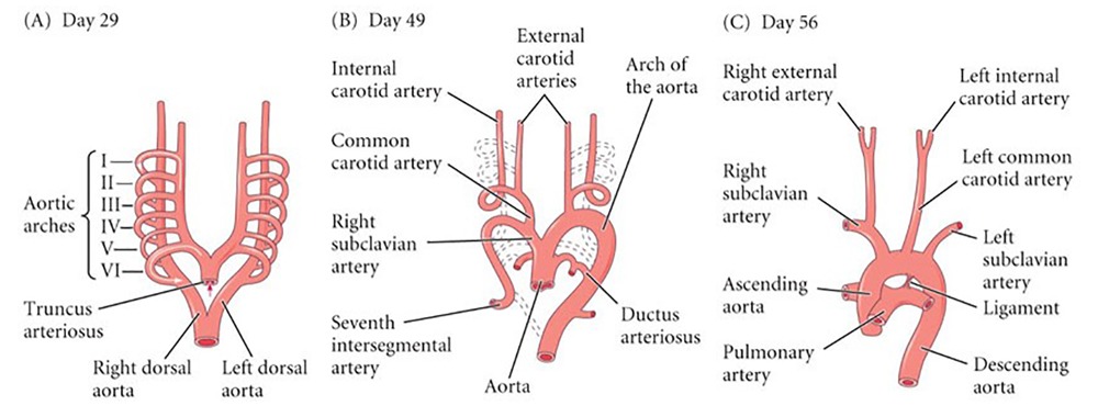
Figure 1: Embryologic development of the six aortic arches into the great vessels of the body.
During fetal development, there are six pairs of aortic arches which initially surround the trachea and esophagus. As fetal development progresses some of the arches will regress while others will go on to form the great vessels. It is when the arches meant to regress instead persist that developmental abnormalities occur. While vascular ring anomalies can arise from the abnormal development of the third, fourth or sixth aortic arches, the most common is the persistence of the fourth right aortic arch (a.k.a. PRAA) which makes up 95% of all cases in dogs and most of the cases in cats.
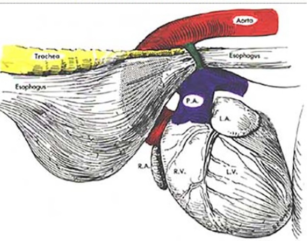
Figure 2: (drawing of vascular ring anomaly) The structure colored green is the vascular ring anomaly (specifically a persistent right aortic arch) causing entrapment and constriction of the esophagus.
As mentioned above, this disorder is typically diagnosed in young animals of 2-6 months of age around the time they are weaned from milk to solid food. Signs observed include failure to grow, thin body condition, voracious appetite, regurgitation, and sometimes even bulging of the neck in the region of the dilated esophagus. Since these animals are at high risk for aspiration pneumonia, other symptoms that may be observed include fever, lethargy, nasal discharge, cough and difficulty breathing. Vascular ring anomalies can be diagnosed with thoracic radiographs (chest x-rays), ideally with use of contrast, which reveal a dilated esophagus in front of the heart. In order to know the specific type of vascular ring anomaly and to aid in surgical planning, a CT scan with angiography needs to be performed.
The treatment of choice is surgical intervention in which the aberrant vessel is ligated and transected alleviating the esophageal constriction. This should be performed as soon as possible after diagnosis in order to reduce the degree of damage to the esophageal muscles and nerves. Prognosis for survival to discharge is 92%, and 87% of cases have good to excellent long-term survival. However, only 30% of cases have complete resolution of clinical signs. Even if the esophagus does not return to normal, as most often some degree of dilation is permanent, episodes of regurgitation become less frequent and body condition greatly improves. A worse prognosis is associated with severity of esophageal dilation and greater time delay to surgery. While medical management can be attempted through elevated feedings of a liquid diet, this is palliative, and the affected animal will continue to have chronic regurgitation, worsening episodes of aspiration pneumonia and failure to thrive due to the inability to consume an appropriate amount of calories. Thus, long term prognosis is poor with this treatment option.
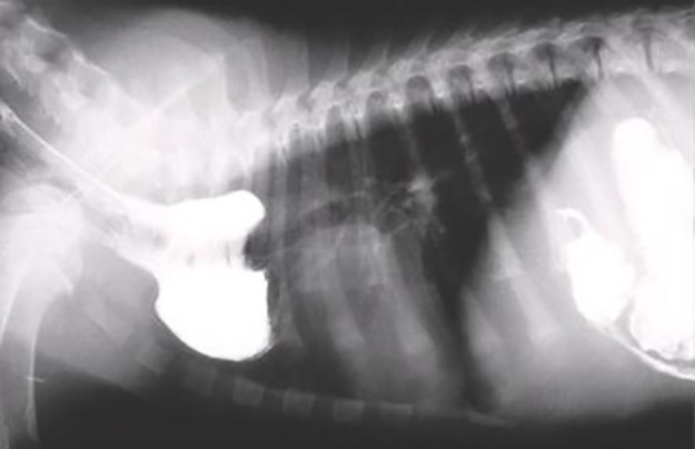
Lateral thoracic radiograph from a dog with chronic regurgitation of food. The radiograph was taken soon after the dog swallowed barium paste. Although the barium paste has passed into the stomach, a portion of it has accumulated in an esophageal dilation cranial to the heart. This appearance is typical for a vascular ring anomaly, most commonly a persistent right aortic arch. Courtesy of Dr. Mark Kittleson © 2005. Figure 3: Barium study of the thorax showing the enlarged esophagus immediately before the heart which is classic for a vascular ring anomaly.
Bibliography:
- Rishniw , Mark, et al. “Vincyclopedia of Diseases: Vascular Ring Anomalies.” VIN, 28 May 2015, vin.com/Members/Associate/Associate.plx?from=GetDzInfo&DiseaseId=17&pid=607.
- Holmberg, D L, and K R Presnell. “Vascular Ring Anomalies: Case Report and Brief Review.” Canadian Veterinary Journal, vol. 20, Mar. 1979, pp. 78–81., ncbi.nlm.nih.gov/pmc/articles/PMC1789496/.
- Plesman, Rhea, et al. “Thoracoscopic Correction of a Congenital Persistent Right Aortic Arch in a Young Cat.” The Canadian Veterinary Journal , vol. 52, Oct. 2011, www.ncbi.nlm.nih.gov/pmc/articles/PMC3174512/.
Images:
- https://www.google.com/imgres?imgurl=https%3A%2F%2Flearninglink.oup.com%2Fstatic%2F5c78d43735e7c000113e2563%2F18-8.jpg&imgrefurl=https%3A%2F%2Flearninglink.oup.com%2Fstatic%2F5c78d43735e7c000113e2563%2Findex.html&tbnid=PGc4LsWK7Sot6M&vet=12ahUKEwim2rDWh6zuAhWZGM0KHUKYBIQQMygEegUIARCyAQ..i&docid=7u7JLkRTHjd8pM&w=840&h=320&q=aortic%20arches%20canine%20embryology&client=firefox-b-1-d&ved=2ahUKEwim2rDWh6zuAhWZGM0KHUKYBIQQMygEegUIARCyAQ
- https://www.vin.com/Members/Associate/Associate.plx?from=GetDzInfo&DiseaseId=17&pid=607
- https://www.vin.com/Members/Associate/Associate.plx?from=GetDzInfo&DiseaseId=17&pid=607